The posterior leg is the largest of the three compartments. This article will describe the anatomy from the inferior view of the skull baseWe will explore the many foramina and projections that enable arteries and nerves to both enter and leave the skullStructure is closely related to function and having an awareness of the location of a structure as well as its function gives an all-round knowledge of the anatomy.
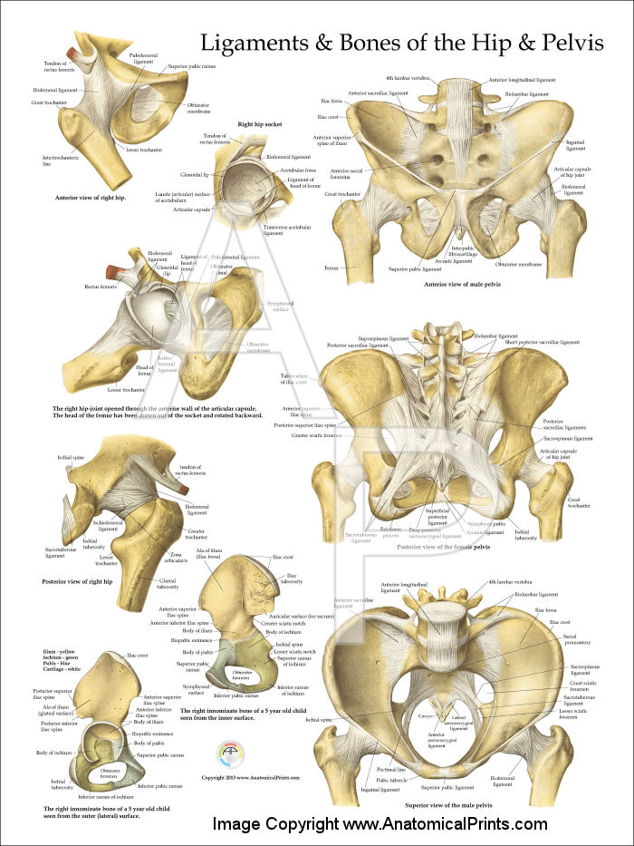
Pelvis And Hip Anatomy Poster
They are innervated by the tibial nerve a terminal branch of the sciatic nerve.
. Pelvic floor weakness is an important problem for many women and basic knowledge of the anatomy of the female pelvic floor is needed to detect it and evaluate its severity. It is attached to the inner surface of each side of the lesser pelvis and these unite to form the greater part of the pelvic floorThe coccygeus muscle completes the pelvic floor which is also called. Collectively the muscles in this area plantarflex and invert the foot.
Pelvic floor weakness may occur with or without prolapse but frequently involves multiple compartments and may require surgical treatment. The pubococcygeus the iliococcygeus and the puborectalis. The levator ani is a broad thin muscle group situated on either side of the pelvisIt is formed from three muscle components.
This is because the rectouterine pouch is the lowest point in the peritoneal cavity when an individual is sitting or standing. In this article we shall look at the attachments actions and innervation of the muscles in the posterior compartment of the leg. The rectouterine pouch also known as the pouch of Douglas or posterior pelvic cul-de-sac can accumulate fluid after a ruptured ectopic pregnancy or an ovarian cyst.
Deep pelvic endometriosis can affect the retrocervical region uterosacral ligaments rectum rectovaginal septum vagina urinary tract and other extraperitoneal pelvic sites. Deep pelvic endometriosis is an important gynecologic disorder that is responsible for severe pelvic pain and is defined as subperitoneal invasion that exceeds 5 mm in depth. Secondarily the ovaries are located posteriorly to the broad ligament so.
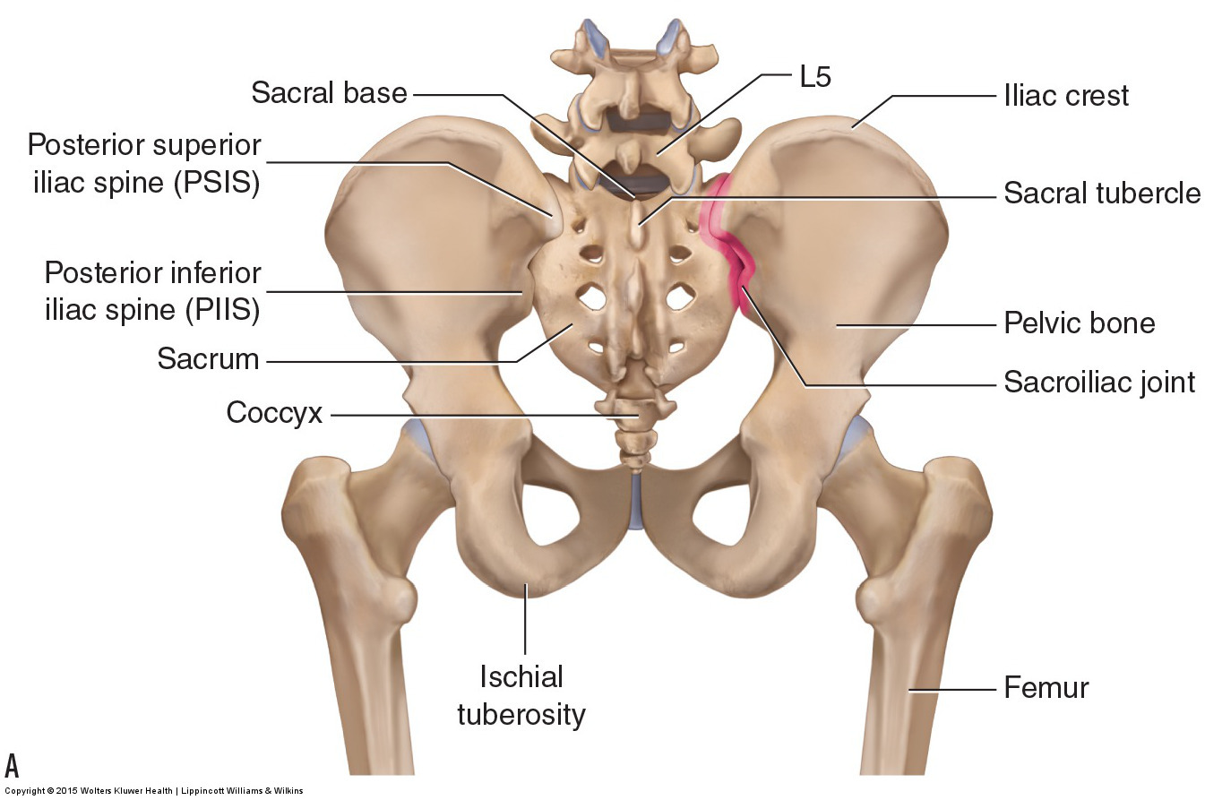
A complete survey of the pelvis is.
Rear View Of Male Pelvis Hip Leg Photograph By Hank Grebe

Three Dimensional Posterior View Of The Pelvis Download Scientific Diagram

The Pelvic Girdle And Pelvis Anatomy And Physiology I

Pelvis Anatomy Recon Orthobullets
Pelvis Anatomy Concise Medical Knowledge
Bones Of The Lumbar Spine And Pelvis
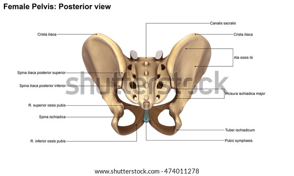
Skeleton Pelvis Posterior View 3d Illustration Stock Illustration 474011278
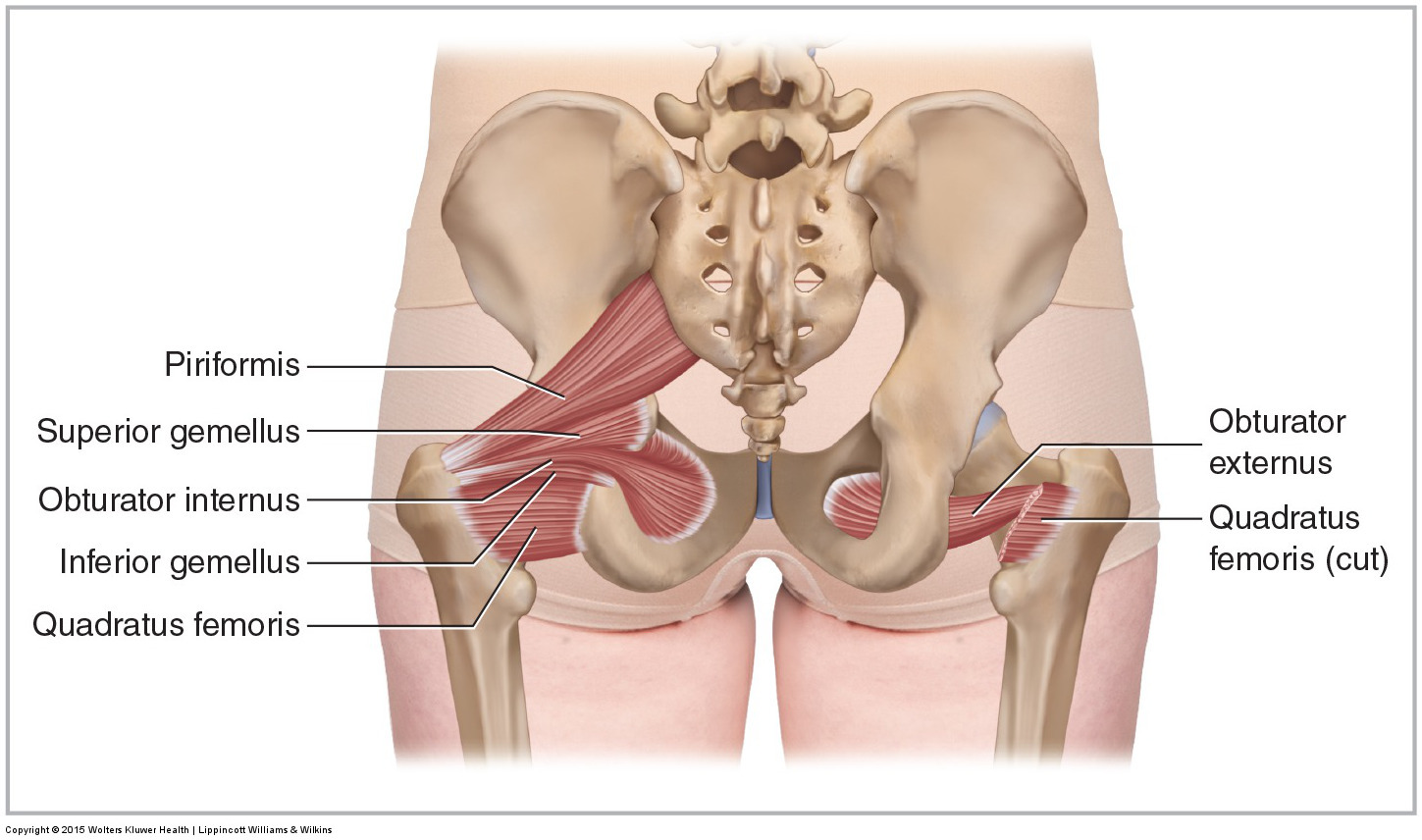
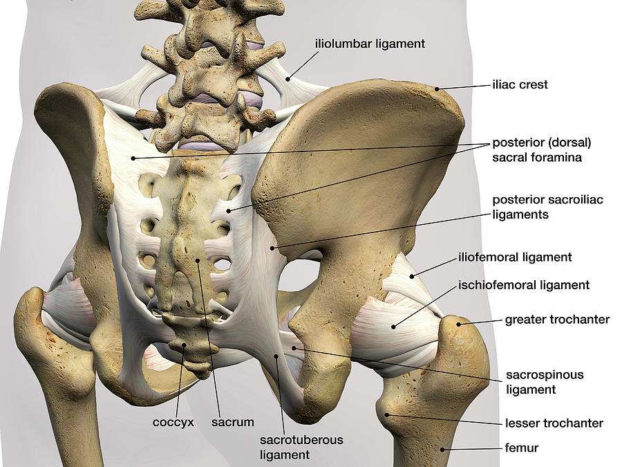
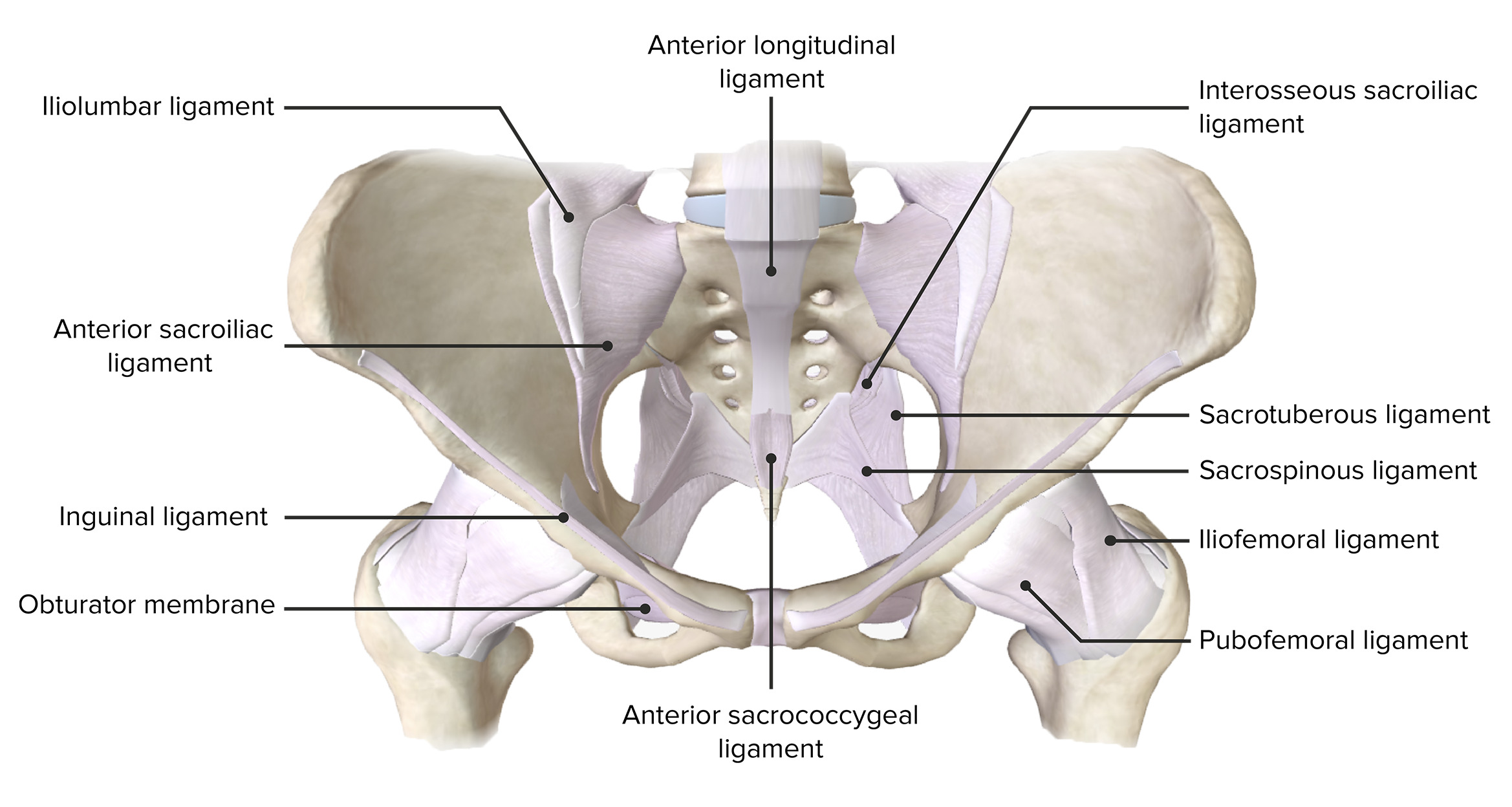
Muscles Of The Pelvis
0 comments
Post a Comment